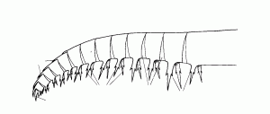
The anatomy of Anomalocaris varies between different species, this is a broad overview of the main characteristic of their basic anatomy, three main taxa have been identified in the Burgess Shale ,
The head of the Anomalocaris is a narrow region compared with the rest of the body and bears a round structure interpreted as a dorsal carapace, it has short flexible dorsolated eye stalks, that were either placed anterior or posterior to the mouth. The mouth known as the oral cone fig 2, was positioned on the underside of the head, and either bilateral or triradial depending on species,
Fossilised Head Fig 1
The oral cone consisted of a central aperture, surrounded by 32 plates, that can be overlapping. consisting of four large plates situated anteriorly, posteriorly and literally 90* apart, or in some species 125* apart. The plates were wedged shaped sclerotized teeth, and some with short spines, The mouth is more rebust than the body and has been well preserved. Fig 2 shows three different species mouth parts of Anomalocaris in the Burgess Shale. The Jaws were segmented and the tooth like prong of the oral cone, continued down the walls of the gullet. The Anomalocaris had a complete digestive system with a advance mid gut region.
Fig2
There were two long tapering frontal appendages on the anterior side of the head as seen in fig 3.These appendages were sclerotized and armed with spines, they were made up of up to 14 podomeres, separated by triangular regions. These appendages could grow up to twenty cm in length. The podomers two to twelve bore a pair of proximal ventral spines that were longer then the height of the associated podomere. the ventrial spine on the second podomere was the longest.
Fig 3
The body of Anomalocaris was elongated and streamline, and could reach a length up to half a meter, its exterior was a smooth surface with or without visible external segmentation. Although segmentation was noted in Anomalocaris canadensis. The body consisted of numerous pairs of lobes, starting from behind the mouth, three pairs of semi-circular flaps and the eleven pairs of triangular lateral lobes, attached to the outer body above each flap and lateral lobe was a multi – structure known as setal blades which were a gill. these lobes were flexible , the third to fifth lobe was the largest part of the body. . the lobes are thought to be composed of muscles surrounding a blood filled cavity . evidence of transverse lines on the anterior part of the flaps are interpreted as strengthening rays or veins .The tail was large and fan-shaped consisting of 3 pairs of prominent dorsolated fins, posterior to the lateral lobes. not a lot is known about the reproductive organs of Anomalocaris, but it is assumed that it would be that of living arthropods today
Dorsal View
References:
Briggs, D.E.G., B.S. Liebermann, J.R. Hendricks, S.L. Halgedahl & R.D. Jarrard. 2008. Middle Cambrian arthropods from Utah. Journal of Paleontology 82(2):238-254.
Butterfield N. J. Leanchoia guts and the interpretation of three dimensional structures in Burgess Shale type fossils. 2001, Paleobiology. 281. 155-171.
Collins D. The evolution of Anomalocaris and its classification in the Arthropods. 1996 Journal of paleeotology 70, 280-293.
Daley A. & Bergstorm. The Oral Cone of Anomalocaris Naturewissenschaften (2012).99 521-504.
Daley, A.C. and Budd, G.E., (2010). Palaeontology, New anomalocaridid appendages from the Burgess Shale, Canada, Volume 53, Issue 4, pages 721–738.
Daley, A. C., Paterson, J. R., Edgecombe, G. D., Garcia-Bellido, D. C., and Jago, J. B. 2013. New Anatomical Information on Anomalocaris from the Cambrian Emu Bay Shale of South Australia and a Reassessment of its Inferred Predatory Habits. The Palaeontological Association. Palaeontology, Vol. 56, Part 5, 2013, pp. 971.
Whittington H. B. & Briggs D,E,G. The largest Cambrian animal, anomalocaris the Burgess Shale.
Yoshiyuki U. Theoretical study on the body form and swimming patterens of Anomalocaris based on hydrodynamic simulation. Journal of theoretical biology (2006) 208, 11-17.


